May 8, 2011
Cranial Nerve III.....
Cranial Nerve III - Oculomotor Nerve
Consists of two components with distinct functions:
Somatic motor
(general somatic efferent) Supplies four of the six extraocular muscles of the eye and the levator palpebrae superioris muscle of the upper eyelid.
Visceral motor
(general visceral efferent) Parasympathetic innervation of the constrictor pupillae and ciliary muscles.
The somatic motor component of CN III plays a major role in controlling the muscles responsible for the precise movement of the eyes for visual tracking or fixation on an object.
The visceral motor component is involved in the pupillary light and accomodation reflexes.
Figure 3-1. Overview of occulomotor nerve components.
Overview of the Somatic Motor Component
There are six extraocular muscles in each orbit.
The somatic motor component of CN III innervates the following four extraocular muscles of the eyes:
Ipsilateral inferior rectus muscle
Ipsilateral inferior oblique muscle
Ipsilateral medial rectus muscle
Contralateral superior rectus muscle
The remaining extraocular muscles, the superior oblique and lateral rectus muscles, are innervated by the trochlear nerve (CN IV) and abducens nerve (CN VI), respectively.
The somatic motor component of CN III also innervates the levator palpebrae superioris muscles bilaterally. These muscles elevate the upper eyelids.
Overview, somatic motor component of CN III.
Actions of the Extraocular Muscles
A knowledge of the origins and points of insertion of the extraocular muscles on the eye relative to the axes of motion of the eye is critical to understanding the actions of these muscles.
Figure 3-3a. Muscles of the orbit.
Medial Rectus Muscle
The medial rectus muscle originates from the tendinous ring of the orbit and inserts on the medial border of the eye. Contraction of this muscles leads to adduction of the eye.
Figure 3-3b. Muscles of adduction.
`Superior Rectus Muscle
The superior rectus muscle originates from the tendinous ring of the orbit and inserts on the superior surface of the eye slightly medial to the eyes vertical axis of rotation. Due to these factors, contraction of the superior rectus results in elevation, intorsion, and adduction of the eye. The primary action of the superior rectus muscle (elevation of the eye) can be isolated by having the patient look laterally and then upwards.
Figure 3-3c. Muscles of elevation.
Actions of the Extraocular Muscles, continued
Inferior Rectus Muscle
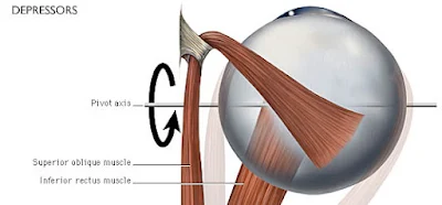
The inferior rectus muscle originates from the tendinous ring of the orbit and inserts on the inferior surface of the eye slightly medial to the eyes vertical axis of rotation. As a result, contraction of the inferior rectus causes depression, extorsion, and adduction of the eye. The primary action of the inferior rectus muscle (depression of the eye) can be isolated by having the patient look laterally and then downwards.
Figure 3-4a. Muscles that depress the eye.
Inferior Oblique Muscle
The inferior oblique muscle originates from the floor of the bony orbit and passes laterally and posteriorly to insert on the posterolateral surface of the eye slightly behind its vertical axis of rotation. Contraction of this muscle results in extorsion (outward rotation about the anterior-posterior axis of rotation), elevation, and abduction. The action of the inferior oblique muscle can be isolated by having the patient look medially and then upwards.
Figure 3-4b. Muscles of abduction.
The actions of the superior oblique and lateral rectus muscles are discussed in the modules on CN IV and CN VI, respectively.
Figure 3-4c. Muscles of medial rotation.
Somatic motor component - origin and central course
The somatic motor component of CN III originates from the oculomotor nucleus located in the rostral midbrain at the level of the superior colliculus.
Like other somatic motor nuclei, the oculomotor nucleus is located near the midline just ventral to the cerebral aqueduct.
In a coronal cross-section of the brainstem the oculomotor nucleus is "V-shaped" and is bordered medially by the Edinger-Westphal nucleus and laterally and inferiorly by the medial longitudinal fasciculus which allows communication between various brainstem nuclei.
Figure 3-5. Somatic motor component - origin and central course.
Fibers leaving the occulomotor nucleus travel ventrally in the tegmentum of the midbrain passing through the red nucleus and medial portion of the cerebral peduncle to emerge in the interpeduncular fossa at the junction of the midbrain and pons.
Cranial Nerve III - Oculomotor Nerve
Somatic motor component - intracranial course
Upon emerging from the brainstem the oculomotor nerve passes between the posterior cerebral and superior cerebellar arteries and pierces the dura mater to enter the cavernous sinus.
The nerve runs along the lateral wall of the cavernous sinus just superior to the trochlear nerve and enters the orbit via the superior orbital fissure.
Figure 3-6. Somatic motor component - intracranial course.
Somatic motor component, final innervation
Within the orbit CN III fibers pass through the tendinous ring of the extraocular muscles and divide into superior and inferior divisions.
The superior division ascends lateral to the optic nerve to innervate the superior rectus and and levator palpebrae superioris muscles on their deep surfaces.
Figure 3-7a. Somatic motor component, final innervation, anterior view.
The inferior division of CN III splits into three branches to innervate the medial rectus and inferior rectus muscles on their ocular surfaces and the inferior oblique muscle on its posterior surface.
Figure 3-7b. Somatic motor component, final innervation.
Somatic motor component, vertical gaze
The exact control of eye movements requires input from integration centers in the brain that coordinate the output from the occulomotor, trochlear, and abducens nuclei which control the six extraocular muscles.
For eye movements in the vertical plane, the superior rectus, inferior rectus, inferior oblique, and superior oblique muscles of the eyes must work precisely together.
The actions of these muscles is coordinated by the vertical gaze center which is thought to be located in the periaqueductal grey matter of the midbrain at the level of the superior colliculus (its location has not yet been positively identified).
The vertical gaze center projects to the oculomotor nuclei which control the superior rectus, inferior rectus and inferior oblique muscles as well as to the trochlear nuclei which control the superior oblique muscles.
The center that controls torsional movements of the eye is probably close to, or the same as, the vertical gaze center since all muscles that elevate or depress the eyes also cause them to rotate about their anterior-posterior axis of motion.
The lateral gaze center is discussed in the abducens nerve (CN VI) module.
Figure 3-8. Vertical gaze center, CN III & CN IV.
Overview of the visceral motor component
Provides parasympathetic innervation of the constrictor pupillae and ciliary muscles of the eye.
The visceral motor component of CN III is involved in the pupillary light and accommodation reflexes.
Figure 3-9. Overview of the visceral motor component.
Visceral motor component, origin and course
The visceral motor component originates from the Edinger-Westphal nucleus located in the rostral midbrain at the level of the superior colliculus.
In a coronal cross-section of the brainstem the Edinger-Westphal nucleus sit within the "V-shaped" oculomotor nuclei just ventral to the cerebral aqueduct.
Preganglionic parasympathetic fibers course ventrally through the midbrain, interpeduncular fossa, cavernous sinus, and superior orbital fissure along with the somatic motor fibers of CN III.
Figure 3-10. Visceral motor component, origin and course.
Visceral motor component, final innervation
Once within the orbit the preganglionic parasympathetic fibers leave the nerve to the inferior oblique muscle to synapse in the ciliary ganglion which lies deep to the superior rectus muscle near the tendinous ring of the extraocular muscles.
Postganglionic fibers exit the ciliary ganglion in the short ciliary nerves which enter the posterior aspect of the eye near the point of exit of the optic nerve.
Within the eye these fibers travel forward between the choroid and sclera to innervate the ciliary muscles (which control the shape and therefore the refractive power of the lens) and the constrictor pupillae muscle of the iris (which constricts the pupil).
Figure 3-11. Visceral motor component, final innervation.
Visceral motor component, pupillary light reflex
Light entering the eye causes signals to be sent to the pretectal region of the midbrain via the optic nerve (CN II).
The pretectal nucleus in turn projects bilaterally to the Edinger-Westphal nucleus.
Preganglionic parasympathetic fibers from each half of the Edinger-Westphal nucleus then project to the ciliary ganglion of the ipsilateral orbit.
Post-ganglionic parasympathetic fibers exit the ciliary ganglion to innervate the constrictor pupillae muscle of the ipsilateral eye.
Due to the bilateral projections from the pretectal nuclei to the Edinger-Westphal nuclei, light shined into one eye produces pupillary constriction in both eyes.
Direct pupillary light reflex - response in the stimulated eye. Consensual pupillary light reflex - response in the opposite eye.
Visceral Motor Component, Accomodation Reflex
Accommodation is an adaptation of the visual apparatus to facilitate near vision. This reflex involves the following:
An increase in the curvature (and therefore the refractive power) of the lens
Pupillary constriction to help sharpen the image on the retina
Convergence of the eyes to fixate on the target object
The accommodation pathway is summarized below:
Fibers from the primary visual cortex project, via the visual association cortex of the occipital lobe, to the superior colliculi and pretectal nuclei.
Axons from the superior colliculi and pretectal nuclei project to both the Edinger-Westphal and oculomotor nuclei.
Signals from the Edinger-Westphal nuclei travel via the ipsilateral oculomotor nerve to reach the ciliary and constrictor pupillae muscles of the eye. Contraction of the ciliary muscle causes the lens to increase its curvature (and thus its refractive power), while contraction of the constrictor pupillae reduces the size of the pupillary aperture.
Signals from the oculomotor nuclei travel via the ipsilateral oculomotor nerve to the medial rectus muscles causing them to contract and resulting in convergence of the eyes on the object of interest.
Action of the ciliary muscle - visceral motor component
The lens of the eye is attached to the ciliary muscle by the suspensory ligaments.
With the ciliary muscle at rest, a certain amount of tension is maintained on the suspensory ligaments keeping the lens relatively flat (low refractive power).
When the ciliary muscle is stimulated to contract the distance from point "A" to point "B" in figure 3-14 is shortened, thus releasing some of the tension on the suspensory ligaments.
With a decrease in tension on the suspensory ligaments the natural elasticity of the lens causes the curvature (and therefore the refractive power) of the lens to increase.
Constriction of the pupil is mediated by the constrictor pupillae muscle.
Lower Motor Neuron (LMN) Lesions
Due to the close proximity of the oculomotor and Edinger-Westphal nuclei and the fact that the fibers of both components travel together all the way to the orbit of the eye, a LMN lesion will most likely affect both components of CN III.
The following collection of signs and symptoms, known as oculomotor ophthalmoplegia, is characteristic of a CN III LMN damage.
Downward, abducted eye on the affected side due to the unopposed actions of the superior oblique and lateral rectus muscles.
Strabismus (the inability to direct both eyes toward the same object) as a result of extraocular muscle paralysis. This leads to diplopia (double vision).
Ptosis (eyelid droop) on the affected side due to inactivation of levator palpebrae superioris muscle and the unopposed action of the orbicularis oculi muscle (innervated by CN VII). The patient may compensate for the ptosis by contracting the muscles of the forehead to raise the eyebrow and lid.
Dilation of the pupil on the affected side due to decreased tone of the constrictor pupillae muscle.
Loss of the accomodation reflex on the affected side.
Because upper motor neuron (UMN) lesions often involve more than one of the cranial nerves, they are discussed in the Eye Movements module.
Clinical correlation - Argyll-Robertson pupil
Argyll-Robertson pupil is caused by damage to cells in the pretectal region of the midbrain. As a result of this damage, signals carried by CN II from the retina are not relayed via the pretectal nucleus on the affected side to the Edinger-Westphal nuclei. This results in a loss of both the direct and consensual pupillary light reflex when light is shined in the eye on the affected side.
Because the accommodation reflex pathway is distinct from the pupillary light reflex pathway the accommodation reflex is unaffected.
Subscribe to:
Post Comments (Atom)


No comments:
Post a Comment