Showing posts with label Diseases. Show all posts

May 10, 2025

Diabetes Mellitus (Type 1 & Type 2) for Nursing & NCLEX Video


Diabetes mellitus (DM) is a chronic metabolic condition that arises from either an absolute or relative lack of insulin, which is an anabolic hormone. Type 1 diabetes, also referred to as insulin-dependent diabetes mellitus (IDDM) or juvenile diabetes, is a persistent condition marked by the body's inability to produce insulin due to the autoimmune destruction of the pancreatic beta cells.

Insulin is generated by the beta cells located in the islets of Langerhans within the pancreas. When these cells are destroyed or otherwise compromised, it leads to the onset of type 1 diabetes (IDDM). Although diabetes mellitus is often perceived as a disease affecting adults, approximately 5% of cases manifest during childhood, typically around the age of 6 or during puberty.


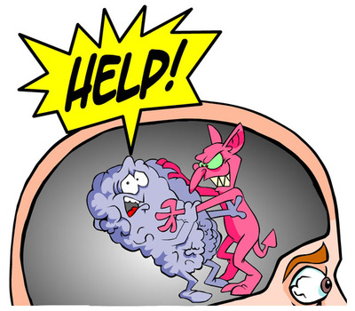
**Pathophysiology of Type 1 Diabetes**

Type 1 diabetes develops when the body does not produce enough insulin, a hormone essential for managing carbohydrates, fats, and proteins. Insulin helps lower blood glucose levels. It allows glucose to enter muscle cells and converts glucose to glycogen (glycogenesis) for storage. Insulin also stops the liver from releasing stored glucose (glycogenolysis) and slows the breakdown of fats into triglycerides, free fatty acids, and ketones. 

When people lack insulin, their blood glucose levels can rise above 200 mg/dL (11 mmol/L), leading to hyperglycemia. This happens because the body cannot use or store glucose properly. 

As a result, the kidneys cannot reabsorb the extra glucose, leading to glycosuria. This process causes more thirst and dehydration. The body also breaks down fats and proteins more, which produces ketones and may lead to weight loss. 

The brain needs glucose for energy. If glucose levels drop below 65 mg/dL (3.2 mmol/L), the body releases hormones like glucagon, cortisol, and epinephrine. This can cause symptoms of hypoglycemia, which can be uncomfortable and alarming. 

Understanding these processes helps us see what people with type 1 diabetes go through. It is important to support those who face these daily challenges.

The glucose level at which symptoms develop varies significantly from person to person and can even change for the same individual over time. This variability is influenced by factors such as the duration of diabetes, the frequency of hypoglycemic episodes, the rate at which blood sugar levels decline, and the overall management of the condition. Understanding these factors is crucial for effective diabetes management and for minimizing the risk of hypoglycemic events.

The overall annual rate of diabetes mellitus is about 24.3 cases for every 100,000 people. Most new cases are type 1 diabetes, with around 15,000 diagnosed each year. However, we’re also seeing a rise in type 2 diabetes among older children, especially within minority groups, with about 3,700 new cases annually. 

A study by Mayer-Davis and colleagues showed that between 2002 and 2012, there was a significant increase in both type 1 and type 2 diabetes among young people in the U.S. After taking into account age, sex, and ethnic backgrounds, they found that type 1 diabetes (in kids aged 0-19 years) had a yearly increase of 1.8%, while type 2 diabetes (in those aged 10-19 years) rose by 4.8% during that time.

Interestingly, the incidence of type 1 diabetes varies quite a bit depending on location. For example, it ranges from just 0.61 cases per 100,000 people in China to 41.4 cases per 100,000 in Finland. Generally, white individuals have the highest rates of type 1 diabetes, while rates are lower among Chinese individuals. It’s important to remember that American whites are 1.5 times more likely to develop type 1 diabetes compared to American blacks or Hispanics. 

Understanding these trends can help us work together to raise awareness and support those affected by diabetes!

In high-incidence areas, older males are at greater risk for type 1 diabetes and may see seasonal variations. Females can be more vulnerable in low-incidence regions. It's essential to consider this diagnosis in infants, as early detection is crucial.


**Clinical Signs and Symptoms of Type 1 Diabetes** 1. **Hyperglycemia**: Sometimes, hyperglycemia can present subtly in children. They may experience general malaise, headaches, or weakness. Increased irritability or sensitivity can also occur, which understandably raises concern for parents and caregivers. ๐Ÿ˜Ÿ
2. **Glycosuria**: The presence of excess glucose in the urine often results in increased frequency and volume of urination (polyuria). This can be particularly challenging at night, leading to nocturia and, in some cases, bedwetting (enuresis) in children who previously had bladder control. ๐ŸŒ™ 3. **Polydipsia**: If your child appears constantly thirsty, it’s not simply a phase; this persistent thirst stems from dehydration induced by osmotic diuresis, which can be distressing for both the child and their caregivers. ๐Ÿ’ง 4. **Polyuria**: A significant rise in urination can be alarming, especially if it results in nighttime accidents. Providing support during this time is crucial, as it might be tough for children to cope with these changes. ๐Ÿ˜Ÿ 5. **Polyphagia**: A notable increase in hunger and food intake may be observed, despite weight loss. This situation is often difficult for parents to witness as it highlights the challenges their child is facing. ๐Ÿฝ️๐Ÿ’” 6. **Weight Loss**: Insulin deficiency can cause noticeable weight loss as the body resorts to breaking down fats and proteins for energy. In younger children, this may appear as failure to thrive and considerable wasting, sometimes before other hyperglycemia symptoms become evident. ⚖️ 7. **Nonspecific Malaise**: Many children may experience a vague sense of malaise before the appearance of any clear symptoms of high blood sugar. This makes it imperative to remain attentive to their overall well-being. ๐Ÿฅบ 8. **Diabetic Ketoacidosis (DKA)**: DKA is a severe condition, and being able to recognize its symptoms is critical. Signs may include drowsiness, dry skin, flushed cheeks, cherry-red lips, a fruity odor on their breath, and deep, labored breathing (Kussmaul breathing). If these symptoms arise, seeking immediate medical assistance is essential. ๐Ÿš‘ Recognizing and understanding these symptoms can be challenging and emotionally draining for families. Approaching these situations with compassion and support is vital in helping children and their families manage the complexities of type 1 diabetes together. ❤️
**Fingerstick Glucose Test**: Children with a family history of diabetes need to have their glucose levels monitored with a fingerstick test. ๐Ÿฉธ **Urine Dipstick Test**: A urine dipstick test can check for ketones in your child's urine, helping you manage their health. ๐Ÿงช **Fasting Blood Sugar (FBS)**: If your child's blood glucose is elevated or if ketonuria is present, a fasting blood sugar test is important. A level of 200 mg/dL or higher may indicate diabetes. ⚖️ **Lipid Profile**: Lipid profiles can show abnormalities at diagnosis due to increased triglycerides. Understanding these changes is key to managing their health. ๐Ÿ“ˆ **Glycated Hemoglobin (HbA1c)**: Monitoring HbA1c levels provides insight into your child's average blood glucose over several weeks, crucial for their long-term management. ๐Ÿ“Š **Microalbuminuria**: This can indicate early signs of nephropathy. Increased albumin excretion is important to track. ๐Ÿ’ง
Medical Management Managing type 1 diabetes in children can feel daunting, but there are supportive strategies available: **Insulin Therapy**: Essential for treatment, insulin doses can be adjusted to maintain normal blood glucose levels. Many children will have two doses daily, helping them lead fulfilling lives. ๐Ÿฝ️ **Diet**: Encouraging a balanced diet high in carbohydrates and fiber yet low in fat supports your child's energy needs. ๐Ÿฅ— **Activity**: Exercise is encouraged, allowing children to participate in sports and activities that benefit their overall well-being. ๐Ÿƒ‍♂️ **Continuous Glucose Monitoring**: This technology helps manage glucose levels effectively, providing peace of mind for families. ๐Ÿ“ฑ Pharmacologic Management Various insulins are available to meet your child's needs: - **Insulin Aspart**, **Insulin Glulisine**, and **Insulin Lispro**: Rapid-acting insulins for flexible dosing. ๐Ÿ’‰ - **Regular Insulin**: Short-acting option for ages 2-18 years. ๐Ÿ•’ - **Insulin NPH**: Intermediate-acting for better control. - **Insulin Glargine** and **Insulin Detemir**: Long-acting insulins that provide stable management. ๐ŸŒ™
- **Insulin Degludec**: Ultra-long-acting insulin for children over 1 year old. ⏳
Nursing Management Caring for a child with diabetes involves: **Assessment**: - Gather information on symptoms and weight changes, allowing your child to share their experiences. ๐Ÿ“‹ - Conduct physical exams to monitor growth, skin health, and glucose levels. ๐Ÿฉบ **Interventions**: - Ensure adequate nutrition based on your child's preferences. ๐Ÿฝ️ - Educate about skin care and recognize signs of hypoglycemia and hyperglycemia to empower both you and your child. ⚠️ Evaluation Progress can be tracked through: - Proper nutrition and skin integrity. ✨ - Infection prevention and regulated glucose levels. ๐Ÿ“ˆ - Supporting your child’s adaptation to diabetes, fostering resilience. ๐ŸŒˆ Documentation Guidelines Accurate documentation supports effective care: - Note findings, intake/output, and cultural beliefs. ๐Ÿ“ - Keep track of care and teaching plans to monitor responses to treatment. ๐Ÿ—‚️ This management approach aims to help children with type 1 diabetes achieve their best health outcomes while providing understanding and support throughout their journey. ๐ŸŒŸ


Additional Information Credits-

Mar 11, 2015

Hepatic Failure News.....

Hepatic failure can result from acute liver injury, causing acute liver failure (ALF) or fulminant hepatic failure (FHF), or progressive chronic liver disease such as cirrhosis. An alteration in hepatocyte functioning affects the liver metabolism, detoxification process, protein synthesis, manufacture of clotting factors, and preservation of immunocompetence. FHF occurs when severe hepatic injury results in encephalopathy and severe coagulopathy within 28 days of the onset of symptoms in patients without a history of chronic liver disease. Liver transplant is the only viable treatment option for patient with FHF. The most commonly identified cause of FHF is drug induced, with acetaminophen the most common culprit, followed by viral hepatitis. Other causes include infection (cytomegalovirus [CMV], adenovirus), metabolic disorders and severe ischemic insult or shock.

Signs and Symptoms

  • Manifestation depends on the complications associated with the liver dysfunction.
  • Patient behavior may range from agitation to frank coma.
  • Evidence of GI bleeding, renal failure, or respiratory distress may also be present.
  • The initial manifestation in FHF is commonly bleeding from coagulopathy.

Physical Examination

Vital signs
  • BP: < 90 mm Hg (with shock)
  • HR: > 120 beats/min (with shock)
  • Temperature may be mildly elevated
  • RR: tachypnea initially progressing to respiratory depression associated with encephalopathy.
Neurologic
  • Mildly confused to coma
  • Personality changes
  • Asterixis
Pulmonary
  • Crackles
  • Labored respirations
Gastrointestinal
  • Hematemesis and melena
  • Ascites
  • Hepatomegaly may be present
  • Splenomegaly may be present
  • Factor hepaticus
  • Diarrhea
Skin
  • Jaundice
  • Ecchymosis and petechiae
  • Pruritus
  • Edema

Acute Care Patient Management

Nursing Diagnosis: Deficient fluid volume related to ascites secondary to hypoalbumineia, bleeding secondary to decreased clotting factors or variceal hemorrhage, and diuretic therapy.
Outcome Criteria
  • BP 90 TO 120 mm Hg
  • Central venous pressure 2 to 6 mm Hg
  • Serum albumin 3.5 to 5 mg/dl
  • Platelet count >50,000/mm3
  • Urine output 30 ml/hr
  • Serum sodium 135 to 145 mEq/L
  • Serum potassium 3.5 to 5 mEq/L
  • Intake approximates output
Patient Monitoring
  1. Obtain pulmonary artery pressure, central venous pressure, and blood pressure until the patient’s condition is stable, then hourly.
  2. Continuously monitor ECG for lethal dysrhythmias that may result from electrolyte and acid-base imbalances.
  3. Monitor fluid volume status. Measure intake and output hourly.
Patient Assessment
  1. Assess hydration status. Note skin turgor on inner thigh or forehead, condition of buccal memranes, and development of edema and crackles.
  2. Assess for signs and symptoms of bleeding.
  3. Measure abdominal girth once each shift to determine progression of ascites.
  4. Assess respiratory status.
Diagnostic Assessment
  1. Review serial serum ammonia, albumin, bilirubin, platelet count, PT, PTT and ALT to evaluate hepatic function.
  2. Review serial serum electrolytes.
  3. Review urine electrolyte, BUN, and creatinine to evaluate renal function.
Patient Management
  1. Administer intravenous crystalloids as ordered.
  2. Administer potassium as ordered. Validate adequate urine output before potassium administration.
  3. Sodium restriction of 0.5 g/day and fluid restriction to 1000 ml/day may be ordered.
  4. Vitamin K or fresh frozen plasma (FFP) may be required to promote the clotting process.
  5. Institute bleeding precautions. Avoid razor blades and use soft-bristled toothbrushes.
  6. Paracentesis may be performed if abdominal distention is severe.
  7. Prepare the patient and family for liver transplant, as indicated.

Jan 12, 2012

  Vasculitis is an inflammation and necrosis of the blood vessels, leading to hemorrhage, ischemia, and infarction.. Treatment of the condition is entirely dependent on the extent of the disease. In many instances, cutaneous vasculitis is a self-limited condition, relieved by leg elevation, avoidance of standing, and therapy with non-steroidal anti-inflammatory drugs (NSAIDs). However, more extensive or severe disease can prove more difficult to manage.


Treatment of vasculitis should begin with a confirmatory diagnosis to eliminate conditions with a similar appearance. A tissue biopsy may confirm diagnosis of cutaneous vasculitis, and can sometimes be used to identify the type of immunoglobin involved. Once a diagnosis is confirmed histologically, other organ systems should be evaluated to identify potential causative factors.


The first and preferred treatment for cutaneous vasculitis should always be avoidance of triggers known to exacerbate the condition, such as excessive standing, infection, or drugs.  For mild recurrent or persistent disease, colchicine and dapsone are first-choice agents.


Severe cutaneous disease requires treatment with systemic corticosteroids or more potent immunosuppression (azathioprine, methotrexate, or cyclophosphamide are typical treatments). A combination of corticosteroids and cyclophosphamide is required therapy for systemic vasculitis, which is associated with a high risk of permanent organ damage or death. Intravenous immunoglobin or plasmapheresis may be useful in the treatment of severe, refractory vasculitis, or in patients who have contraindications to traditional immunosuppression.


New biologic therapies that act via cytokine blockade or lymphocyte depletion, such as the tumor necrosis factor-ฮฑ inhibitor infliximab and the anti-B-cell antibody rituximab, respectively, appear to offer some benefit in certain settings, such as connective tissue disease and anti-neutrophil cytoplasmic antibody-associated vasculitis.


This is article 3 in a series of four articles pertaining to vasculitis.  To read the additional articles click title link, or  click here.


.
Vasculitis is the inflammation of blood vessels. There are many types of vasculitis. Diseases in which vasculitis is a primary process are called primary systemic vasculitides. Vasculitis may also occur as a secondary feature in other rheumatic diseases and syndromes

WHAT YOU NEED TO KNOW ABOUT VASCULITIS

What is vasculitis?
Vasculitis is a general term that refers to the inflammation of blood vessels. When blood vessels become inflamed, they can only react in limited ways. They may become weakened, stretch and increase in size, or become narrow – even to the point of closing off entirely.


What are the consequences of vasculitis?
In an extreme situation, when a segment of a blood vessel becomes weakened, it may then stretch and bulge (called an “aneurysm”). The wall of the blood vessel can become so weak that it ruptures and bleeds. Fortunately, this is a very rare event.


If a blood vessel becomes inflamed and narrowed, blood supply to that area may be partially or completely eliminated. If collateral blood vessels (thought of as alternate routes of blood supply) are not available in sufficient quantity to carry the blood to such sites, the tissue supplied by the affected blood vessels will die. This is called infarction.

Because vasculitis can occur in any part of the body, any tissue or organ can be at risk.


Who is affected by vasculitis?
Vasculitis can affect people of all ages from childhood to adulthood. There are some types of vasculitis that occur in certain age groups more than others.


What are the causes of vasculitis?
Vasculitis may occur secondary to an identified underlying disease or trigger. Occasionally, an allergic reaction to a medicine may trigger vasculitis. Vasculitis can sometimes develop in conjunction with an infection. Usually in these cases, the infection causes an abnormal response in the person’s immune system, damaging the blood vessels. Viral hepatitis (a type of liver infection), is a specific infection that can be associated with vasculitis. Vasculitis may also be related to other diseases of the immune system that the patient had for months or years. For example, vasculitis could be a complication of rheumatoid arthritis, systemic lupus erythematosus, or Sjรถgren’s syndrome.


In many cases though, the causes of vasculitis are not known. These diseases are collectively sometimes referred to under the broad heading of primary forms of vasculitis. In such settings, the appearance and location of the vasculitis often behaves in a distinct way allowing it to be diagnosed as a unique type of vasculitis and is given a specific name.


What are the types of primary vasculitis?
There are many types of primary vasculitis including disease entities such as Wegener’s granulomatosis, microscopic polyangiitis, Henoch-Schรถnlein purpura, polyarteritis nodosa, Kawasaki disease, giant cell arteritis, Takayasu’s arteritis, and Behรงet’s disease. Some are named after doctors (Wegener, Takayasu, Kawasaki) who were among those to provide the best original descriptions of the illness or are named based on features seen on biopsies (giant cell arteritis, angiitis, arteritis nodosa) of affected tissues or blood vessels. Although most of these are systemic (or generalized) vasculitides where the vasculitis may affect many organ systems at the same time, they often differ a great deal among each other. Some of the primary systemic vasculitic diseases may be quite mild and require little or even no treatment. Other forms may be severe, affecting critical organs and, if left untreated, may lead to death within days or months.

Some forms of primary vasculitis may be restricted in their location to certain organs (these are called isolated forms of vasculitis). Examples include vasculitis that only occurs either in the skin, eye, brain (isolated CNS vasculitis) or certain internal organs.


What are the symptoms of vasculitis?
Because any organ system may be involved, an enormous number of symptoms are possible. If the skin is involved, there may be a rash. If nerves suffer loss of blood supply, there may initially be an abnormal sensation followed by a loss of sensation. Vasculitis in the brain may cause a stroke, or in the heart may result in a heart attack. Kidney inflammation usually is not associated with symptoms and is detected by the doctor by examination of the urine. This is important to recognize as inflammation in the kidneys can lead to kidney failure unless promptly detected.


Sometimes the symptoms are nonspecific. When inflammation is present in the body, we tend to respond in ways that tell us that we are not well, but those responses may not be unique to vasculitis at all. For example, along with the symptoms mentioned previously, a person with vasculitis may also have a fever or experience loss of appetite, weight loss and loss of energy.


How is vasculitis treated?
Treatment depends entirely upon the diagnosis, the organs that are affected, and the severity of the vasculitis. When vasculitis represents an allergic reaction, it may be “self limiting,” or will go away on its own and not require treatment. There are other instances also where minimal to no treatment is required and the person can be closely observed.


In instances where critical organs such as the lungs, brain or kidneys are involved, the outlook is less positive and aggressive and timely treatment is necessary. For most forms of systemic vasculitis, treatment generally includes corticosteroid medications (prednisone is the most commonly prescribed).


For some forms of vasculitis, treatment must also include another immunosuppressive medication used in combination with the prednisone. Some of these medications are chemotherapy agents like those used to treat cancer, but are given in doses considerably lower than people with cancer may receive. The goal of this type of chemotherapy is to suppress the abnormal immune response that has led to blood vessel damage.


What is the outlook for people with vasculitis?
The outlook for a person who has vasculitis will vary with the type of vasculitis that is present, what organs are being affected, how severe the vasculitis is, and how the person responds to treatment. Knowing the type of vasculitis allows the doctor to predict the likelihood of illness severity and outcome.


Prior to the time of available treatment, people with severe vasculitis may have had anticipated survival of only weeks to months. However, today with proper treatment, normal life spans are possible. The success of therapy is related to prompt diagnosis, aggressive treatment and careful follow-up to be sure that side effects from medications do not develop.


Once vasculitis is under control (often referred to as “remission”), medications may be cautiously withdrawn, with the hope that the patient will sustain a long remission, independent of treatment. Because some forms of vasculitis can recur (referred to as a “relapse”) after a period of remission, it is very important for patients with vasculitis to remain under the care of a knowledgeable physician.

Jan 9, 2012

Diabetes Animation...Maintaining Blood Glucose Homeostasis.....

Adherence to Post MI Medications

Stroke Risk Associated With Diet Quality, Energy Intake Overall.....


Clinical Context...

Undernutrition before birth, as well as in infancy, childhood, and adulthood, is associated with an increased risk for stroke in later life. However, the mechanism of increased stroke risk is poorly understood.
In addition, overnutrition also increases stroke risk. The likely mechanism is by hastening the onset of obesity, hypertension, hyperlipidemia, and diabetes. The objective of this review by Hankey was to describe recent evidence regarding the effects of nutrition on stroke risk.

Study Synoposis and Perspective

The overall quality of a person’s diet and the balance between caloric intake and caloric expenditure appear to be more important determinants of stroke risk than the actual foods and nutrients consumed, according to a new review published in the January 2012 special issue of the Lancet Neurology.
In general, the 2 biggest threats to health and risk of stroke are overeating and excess salt, author Graeme J. Hankey, from Royal Perth Hospital, Perth, Western Australia, writes.

"These behaviors are a normal response by people to an abnormal environment," Dr. Hankey notes. "Our living environments have become more conducive to consumption of energy and less conducive to expenditure of energy in developed and increasingly in developing regions."
He writes that between 1970 and 2008, the incidence of stroke in high-income countries fell by 42%, probably as a result of increased public awareness about the dangers of high blood pressure, high cholesterol, and cigarette smoking.

But in poorer countries, the incidence of stroke increased by more than 100% during this period. This rise coincided with food and lifestyle changes associated with industrialization and urbanization.


Small Number of Trials
In his review, Dr. Hankey examines the evidence linking nutrition and diet to the risk of stroke. He searched PubMed articles published in English from 1970 to October 2011, using a variety of search terms.
The review included a small number of randomized trials and large observational and epidemiological studies.
He found that the findings from these studies were diverse, owing to the fact that most were epidemiological and therefore "prone to substantial methodological challenges of bias, confounding, and measurement error."
For example, one observational study found that a high intake of a healthy diet was associated with an increased risk of stroke, whereas another observational study found just the opposite.
Dr. Hankey's review also revealed the following effects of individual foods and beverages on the risk of stroke:

  • Fish: 3 servings a day associated with a 6% lower risk of stroke
  • Fruits and vegetables: >5 servings a day associated with a 26% lower risk of stroke
  • Meat: Each daily serving associated with a 24% increased risk of stroke
  • Reduced-fat milk: associated with a lower risk of stroke vs full-fat milk
  • Chocolate: High consumption associated with a 29% lower risk of stroke
  • Coffee: 3 to 4 cups per day associated with a 17% lower risk of stroke
  • Tea: ≥3 cups per day associated with a 21% lower risk of stroke
  • Sugar-sweetened beverages: high intake associated with increased obesity, diabetes, metabolic syndrome, and coronary heart disease
  • Whole grains: high intake associated with a 21% lower incidence of cardiovascular events
  • Rice: intake not associated with risk of stroke
  •  
"Further research is needed to improve the quality of evidence relating to the association of many nutrients, foods, and dietary patterns with stroke risk," Dr. Hankey suggests.
This requires adequately powered, large randomized trials with carefully described patient populations and interventions. The outcomes of such trials should also distinguish first-ever and recurrent stroke, as well as ischemic and hemorrhagic stroke, and their different etiologies.
Dr. Hankey also suggests that population-wide salt reduction programs led by governments and with industry cooperation could be highly cost effective.
"In the USA, modest, population-wide reductions in dietary salt of up to 3 g per day…are projected to reduce the annual number of new cases of stroke by 32,000 to 66,000, similar to the benefits of population-wide reductions in tobacco use, obesity, and cholesterol levels," he writes.

Dr. Hankey told Medscape Medical News that he hopes his study will stimulate conclusive studies "by means of large, randomized controlled trials of the effect of dietary nutrients, foods, and patterns on stroke and its pathological and etiological subtypes."
The current evidence associating dietary nutrients, foods, and patterns with stroke is generally suboptimal for providing reliable conclusions about causality, he reiterated.
"The effect of only a few supplements on stroke risk have been studied by means of large, randomized controlled trials, and all of those have shown no benefit of antioxidant vitamins, B-vitamins, and calcium supplements in preventing stroke, despite large, more suboptimal, epidemiological studies claiming they should be beneficial," he said.


Best Diet Still Uncertain
Commenting on this study for Medscape Medical News, Eugene Storozynsky, MD, a cardiologist from the University of Rochester Medical Center, Rochester, New York, said that he agrees with Dr. Hankey's conclusions.

"This is a good review of the literature as to what role diets and their components would have on reducing stroke, and what it tells us is that there still is a lot of uncertainty about the best diet. One study suggests benefit of a certain food or strategy, the other may come to the opposite conclusion," Dr. Storozynsky said.
The take-home message here is as Dr. Hankey notes, Dr. Storozynsky continued. "We need rigorously done randomized controlled trials to really monitor the long-term effects of diets and nutrients, rather than observation studies that look at individual factors, to see whether or not there is any benefit to adopting certain strategies."


Dr. Storozynsky also noted what he considers to be an important omission from Dr. Hankey's review.
"This review did not look at the effect of exercise and how it affects nutrition or stroke results. Does routine daily exercise mitigate the effect of intermittent poor eating? Exercise definitely needs to be part of the equation," he said.


Helmi L. Lutsep, MD, from the Oregon Stroke Center at Oregon Health Science University in Portland, added, "My quick read of the article suggests that it is a very nicely written, comprehensive review of our current knowledge regarding nutrition and stroke."
Dr. Lutsep, who is also a spokesperson for the American Academy of Neurology, added, "While I haven't seen all of these pieces summarized in one place before, I don't think that the article provides novel observations."

Dr. Hankey reports that he was the principal investigator of the VITAmins TO Prevent Stroke (VITATOPS) trial and has financial relationships with Sanofi-Aventis, Johnson & Johnson, Bristol-Myers Squibb, Boehringer Ingelheim, Bayer, and Pfizer Australia. Dr. Storozynsky and Dr. Lutsep have disclosed no relevant financial relationships. 

 
Lancet Neurol. 2012;11:66-81. Abstract
The National Stroke Association provides a comprehensive discussion for patients about stroke prevention.

Study Highlights


  • On the basis of a literature review, the reviewer noted the following findings:
    • Vitamin A supplementation increases all-cause mortality risk.
    • ฮฒ-carotene supplementation increases cardiovascular and all-cause mortality risks and does not prevent stroke.
    • Vitamin C supplementation does not prevent stroke.
    • Vitamin E supplementation increases all-cause mortality risk and does not prevent stroke.
    • Although folic acid supplementation does not prevent stroke in populations with high folate intake, deficiency in regions of low folate intake may be a causal and treatable risk factor for stroke.
    • Vitamin D deficiency is associated with hypertension, cardiovascular disease, and stroke, but to date, supplementation has not been shown to prevent cardiovascular events. Randomized trials are underway.
    • Salt supplementation by 5 g per day is linked to a 23% increase in stroke risk (95% confidence interval [CI], 6 - 43).
    • Although decreasing salt intake has not been shown to reduce stroke risk, lowering consumption by 2 g per day is associated with a 20% decrease in cardiovascular events (95% CI, 1 - 36), and lower salt intake is also linked to blood pressure reduction.
    • Potassium supplementation by 1 g per day is associated with an 11% reduction in the risk for stroke (95% CI, 3 - 17), but supplementation is not proven to prevent stroke.
    • Potassium supplementation by 0.8 g per day is associated with a decrease in blood pressure by 5/3 mm Hg.
    • Calcium supplementation exceeding 0.5 g per day is linked to a 31% increase in the risk for myocardial infarction (95% CI, 2 - 67), does not prevent stroke, and may actually increase stroke risk.
    • High intake of total fat, trans fats, and saturated fats is not associated with an increased risk for stroke, and reduced total fat intake does not lower stroke risk.
    • High intake of plant n-3 polyunsaturated fats is associated with a reduced risk for stroke.
    • Marine n-3 polyunsaturated fat supplementation lowers cardiovascular events and death by 8% (95% CI, 1 - 15), but in a randomized trial, it did not reduce stroke risk.
    • High intake of carbohydrates with high glycemic index and glycemic load is associated with increased blood glucose levels, body weight, and stroke mortality.
    • High fiber intake is linked to lower blood pressure, blood glucose levels, and low-density lipoprotein cholesterol levels.
    • High protein intake is not associated with stroke risk.
    • High intake of a healthy diet was linked to an increased risk for stroke in one observational study and a reduced risk for stroke in another observational study.
    • High intake of an unhealthy diet was linked to an increased risk for stroke and a population-attributable risk for stroke of 19% (99% CI, 11 - 30).
    • In women, a prudent diet or Dietary Approaches to Stop Hypertension (DASH)–style diet is associated with a lower risk for stroke, and a Western diet is linked with a higher risk for stroke.
    • In women, the Mediterranean diet is associated with lower risks for stroke, cardiovascular disease, cardiovascular mortality, and all-cause mortality.
    • The effects of a vegetarian diet and a Japanese diet on stroke risk are unknown.
    •  
    •  

Clinical Implications


  • Reliable evidence from randomized trials suggests that dietary supplementation with antioxidant vitamins, B vitamins, and calcium does not reduce the risk for stroke. However, methodologic limitations of studies to date preclude firm conclusions regarding the effect of many other nutrients on stroke risk.
  • Less reliable evidence suggests that stroke can be prevented by prudent diets; Mediterranean or DASH-type diets; diets that are low in salt and added sugars and high in potassium; and diets that meet, but do not exceed, energy requirements. The overall quality of dietary pattern and the balance between energy intake and expenditure seem to affect stroke risk to a greater extent than individual nutrients and foods.

Borrowed from- Medscape Education Clinical Briefs

Study in monkeys raises hope for HIV .......vaccines

Study in monkeys raises hope for HIV vaccines

Click the Above link to read the Article....

Dec 23, 2011

Glucose Testing....

Glucose


Also known as: Blood sugar; Fasting blood sugar; FBS; Fasting blood glucose; FBG; Fasting plasma glucose; FPG; Blood glucose; Oral Glucose Tolerance Test; OGTT; GTT; Urine glucose
Formal name: Blood Glucose; Urine Glucose

At a Glance

Why Get Tested?

To determine if your blood glucose level is within a healthy range; to screen for, diagnose, and monitor high blood glucose (hyperglycemia) or low blood glucose (hypoglycemia), diabetes, and pre-diabetes; to check for glucose in your urine

When to Get Tested?

Blood glucose: as part of a regular physical, when you have symptoms suggesting hyperglycemia or hypoglycemia, and during pregnancy; if you are diabetic, self-checks up to several times a day to monitor blood glucose levels
Urine glucose: usually as part of a urinalysis

Sample Required?

A blood sample drawn from a vein in your arm or a drop of blood from a skin prick; sometimes a random urine sample is used. Some diabetic patients may use a continuous glucose monitor, which is a small sensor wire inserted beneath the skin of the abdomen that measures blood glucose every five minutes.

Test Preparation Needed?

In general, it is recommended that you fast - nothing to eat or drink except water - 8 hours before having a blood glucose test. In persons with diabetes, glucose levels are often checked both while fasting and after meals to provide the best control of diabetes. For random, timed, and post-meal glucose tests, follow your doctor's instructions. 

The Test Sample

What is being tested?

This test measures the amount of glucose in the blood or urine. Glucose is the primary energy source for the body’s cells and the only energy source for the brain and nervous system. A steady supply must be available for use, and a relatively constant level of glucose must be maintained in the blood.
During digestion, fruits, vegetables, breads and other carbohydrates are broken down into glucose (and other nutrients); they are absorbed by the small intestine and circulated throughout the body. Using glucose for energy production depends on insulin, a hormone produced by the pancreas. Insulin facilitates transport of glucose into the body's cells and directs the liver to store excess energy as glycogen for short-term storage and/or as triglycerides in adipose (fat) cells.
Normally, blood glucose rises slightly after a meal and insulin is released by the pancreas into the blood in response, with the amount corresponding to the size and content of the meal. As glucose moves into the cells and is metabolized, the level in the blood drops and the pancreas responds by slowing, then stopping the release of insulin.
If the blood glucose level drops too low, such as might occur in between meals or after a strenuous workout, glucagon (another pancreatic hormone) is secreted to induce the liver to turn some glycogen back into glucose, raising the blood glucose level. If the glucose/insulin feedback mechanism is working properly, the amount of glucose in the blood remains fairly stable. If the balance is disrupted and the glucose level in the blood rises, then the body tries to restore the balance, both by increasing insulin production and by eliminating excess glucose in the urine.

There are a few different conditions that may disrupt the balance between glucose and the pancreatic hormones, resulting in high or low blood glucose. The most common cause is diabetes. Diabetes is a group of disorders associated with insufficient insulin production and/or a resistance to insulin. People with untreated diabetes are not able to process and use glucose normally. Those who are not able to produce enough insulin to process glucose are diagnosed as having type1 diabetes while people who are resistant to insulin have type 2. Either type of diabetic may have acute and/or chronically increased blood glucose levels.
Severe, acute high blood glucose (hyperglycemia) or low blood glucose (hypoglycemia) can be life-threatening, causing organ failure, brain damage, coma, and, in extreme cases, death. Chronically high blood glucose levels can cause progressive damage to body organs such as the kidneys, eyes, heart and blood vessels, and nerves. Chronic hypoglycemia can lead to brain and nerve damage.
Some women may develop hyperglycemia during pregnancy, which is termed gestational diabetes. If untreated, this can cause these mothers to give birth to large babies who may have low glucose levels. Women who have had gestational diabetes may or may not go on to develop diabetes.

How is the sample collected for testing?

A blood sample is obtained by inserting a needle into a vein in the arm or a drop of blood is taken by pricking the skin, typically on a finger, with a small, pointed lancet (fingerstick). Sometimes, a random urine sample is collected. Some diabetics may use a continuous glucose monitor, which is a small sensor wire inserted beneath the skin of the abdomen and held in place with an adhesive patch. The sensor measures blood glucose levels every five minutes and sends the results to a device that is attached to the person's clothing. A digital readout on the device lets the person know the blood glucose level in real time.

Is any test preparation needed to ensure the quality of the sample?

For screening purposes, fasting is generally recommended - nothing to eat or drink except water - at least 8 hours before a blood glucose test. Those who have been diagnosed with diabetes and are monitoring glucose levels are often tested both while fasting and after meals. For random and timed tests, follow the doctor's instructions. A glucose tolerance test requires that you fast for the first blood sample and then drink a liquid containing a specified amount of glucose. Subsequent blood samples are drawn at specified times.

The Test

Common Questions

Article Sources

(Revised 2011 February). Know your Blood Sugar Numbers. National Diabetes Education Program [On-line information]. PDF available for download at http://ndep.nih.gov/media/knownumbers_eng.pdf through http://ndep.nih.gov. Accessed May 2011.
Olatunbosun, S. (Updated 2011 April 19). Glucose Intolerance. Medscape Reference [On-line information]. Available online at http://emedicine.medscape.com/article/119020-overview through http://emedicine.medscape.com. Accessed May 2011.
Dugdale, D. (Updated 2010 May 23). Glucose test – blood. MedlinePlus Medical Encyclopedia [On-line information]. Available online at http://www.nlm.nih.gov/medlineplus/ency/article/003482.htm. Accessed May 2011.