Showing posts with label Cranial Nerves. Show all posts
Showing posts with label Cranial Nerves. Show all posts
Feb 6, 2013
Labels:
Anatomy,
Brain,
Cranial Nerves,
illustrations. anatomy. nerves,
Pathophysiology,
Physical Assessment,
Physiology
May 23, 2011
Branches of facial nerve: Temporal, Zygomatic, Buccal, Mandibular, Cervical (Ten Zebras Bought My Car).....
May 9, 2011
Hypoglossal Nerve (CN XII) ............
Also known as CN XII, the hypoglossal nerve is the twelfth of twelve paired cranial nerves. The CN XII innervates the muscles of the tongue. It is a somatomotor nerve whose fibers originate in the hypoglossal nucleus neurons situated in the dorsal medulla oblongata of the brainstem. The hypoglossal nucleus nerve cells send axons that exit as rootlets that emerge in the ventrolateral sulcus of the medulla between the olive and pyramid. Then, the rootlets come together to make up the hypoglossal nerve, exiting the cranium through the hypoglossal canal.
As the CN XII comes out of the hypoglossal canal, it gives off a small meningeal branch, picking up a branch from the anterior ramus of C1. Then, it spirals behind the vagus nerve and passes between the internal carotid artery and internal jugular vein lying on the carotid sheath. After passing deep to the posterior belly of the digastric muscle, the hypoglossal nerve passes to the submandibular region to enter the tongue.
Labels:
Anatomy,
Cranial Nerves,
Physiology
Accessory Nerve (CN XI) .............
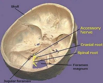
Although it originates in the central nervous system, the spinal accessory nerve begins outside the skull rather than inside, with its axonal fibers arising from neurons located in the upper spinal cord, near the medulla oblongata. These fibers coalesce to form the spinal accessory nerve, which enters the skull through the foramen magnum, the large opening at the base of the skull. Then the nerve runs along the inner wall of the skull towards the jugular foramen, through which it exits the skull together with the glossopharyngeal (CN IX) and vagus nerves (CN X). Thus, the accessory nerve is notable for being the only cranial nerve to both enter and exit the skull.
Labels:
Anatomy,
Cranial Nerves,
Physiology
Vagus Nerve (CN X) .........
The vagus nerve also supplies motor parasympathetic fibers to all the organs except the suprarenal glands, from the neck down to the second segment of the transverse colon. This means that the vagus nerve is responsible for such varied tasks as heart rate, gastrointestinal peristalsis, sweating, and quite a few muscle movements in the mouth, including speech (via the recurrent laryngeal nerve) and keeping the larynx open for breathing, via action of the posterior cricoarytenoid muscle, the only abductor of the vocal folds. It also has some afferent fibers that innervate the inner (canal) portion of the outer ear, via the Auricular branch, which also known as Alderman's nerve, and part of the meninges. This explains why a person may cough when tickled on their ear (such as when trying to remove ear wax with a cotton swab).
Labels:
Anatomy,
Cranial Nerves,
Physiology
Subscribe to:
Comments (Atom)


