May 8, 2011
Cranial Nerve IV - Trochlear Nerve Review.....
Cranial Nerve IV - Trochlear Nerve
Overview
The trochlear nerve has only a somatic motor component:
Somatic motor
(general somatic efferent) Somatic motor innervates the superior oblique muscle of the contralateral orbit.
The superior oblique muscle is one of the six extraocular muscles responsible for the precise movement of the eye for visual tracking or fixation on an object.
See the occulomotor nerve (CN III) chapter for a discussion of eye movements and the interaction between the three nuclei and nerves that innervate the extraocular muscles.
Figure 4-1. Anatomic overview of the trochlear nerve.
The trochlear nerve innervates the superior oblique muscle of the contralateral orbit.
Contraction of the superior oblique muscle depresses, intorts (rotates inward), and abducts the eye:
Figure 4-2. Superior oblique muscle.
Origin and central course
The fibers of the trochlear nerve originate from the trochlear nucleus located in the tegmentum of the midbrain at the level of the inferior colliculus.
The nucleus is located just ventral to the cerebral aqueduct. It is readily identifiable by its close association with the myelinated medial longitudinal fasciculus that allows communication between various brainstem nuclei.
Fibers leaving the trochlear nucleus travel dorsally to wrap around the cerebral aqueduct.
All fibers of the two trochlear nerves decussate (i.e. cross) in the superior medullary velum and exit the dorsal surface of the brainstem just below the contralateral inferior colliculus.
Figure 4-3. Origin and central course.
Intracranial course
Intracranial course and final innervation
From the cavernous sinus the trochlear nerve enters the orbit through the superior orbital fissure.
CN IV does not pass through the tendinous ring of the extraocular muscles, rather it passes above the ring.
The trochlear nerve then crosses medially along the roof of the orbit above the levator palpebrae and superior rectus muscles to innervate the superior oblique muscle along its proximal one-third:
Figure 4-5. Final innervation of trochlear nerve.
Upon emerging from the dorsal surface of the brainstem the trochlear nerve curves around the brainstem in the subarachnoid space and emerges between the posterior cerebral and superior cerebellar arteries (along with CN III fibers).
The trochlear nerve then enters and runs along the lateral wall of the cavernous sinus with CNS III, V, and VI.
Figure 4-4. Intracranial course.
Clinical Correlation
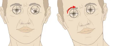
The superior oblique muscle normally depresses, intorts, and abducts the eye (fig. 4-6a). Damage to the trochlear nerve will present as:
Extorsion (outward rotation) of the affected eye due to the unopposed action of the inferior oblique muscle.
Vertical diplopia (double vision) due to the extorted eye.
Weakness of downward gaze most noticeable on medially-directed eye. This is often reported as difficulty in descending stairs.
Figure 4-6a. Normal action of the superior oblique muscle.
Extortion (outward rotation) of the affected eye due to the unopposed action of the inferior oblique muscle (Fig. 4-6b). Vertical diplopia (double vision) due to the extorted eye. Weakness of downward gaze most noticeable on medially directed eye. This is often reported as difficulty in descending stairs. Head tilt (Fig. 4-6b): patient will often tilt his head opposite the side of the affected eye in an attempt to compensate for the outwardly rotated eye.
Due to its long peripheral course around the midbrain CN IV is particularly susceptible to head trauma.
Unique Features
The trochlear nerve has several features that make it unique from the other cranial nerves:
Is the only nerve to exit from the dorsal surface of the brain.
Is the only nerve in which all the lower motor neuron fibers decussate.
Has the longest intracranial course.
Has the smallest number of axons......
Subscribe to:
Post Comments (Atom)


No comments:
Post a Comment