Branchial Motor Component
The largest component of the facial nerve.
Provides voluntary control of the muscles of facial expression (including buccinator, occipitalis and platysma muscles), as well as the posterior belly of the digastric, stylohyoid and stapedius muscles.
Note the branchial motor components of the facial nerve:
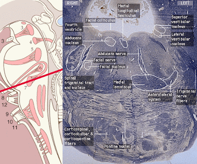
The branchial motor component originates from the motor nucleus of CN VII in the caudal pons.
Fibers leaving the motor nucleus of CN VII initially travel medially and dorsally to loop around the ipsilateral abducens nucleus (CN VI) producing a slight bulge in the floor of the fourth ventricle - the facial colliculus.
Fibers then course so as to exit the ventrolateral aspect of the brainstem at the caudal border of the pons in conjunction with the nervus intermedius components of CN VII.
Figure 7-3. Brainstem section: origin and central course of the branchial motor components of the facial nerve.
Intracranial course
Upon emerging from the ventrolateral aspect of the caudal border of the pons, all of the components of CN VII enter the internal auditory meatus along with the fibers of CN VIII (vestibulocochlear nerve).
The fibers of CN VII pass through the facial canal in the petrous portion of the temporal bone. The course of the fibers is along the roof of the vestibule of the inner ear, just posterior to the cochlea.
| Figure 7-4. Intracranial course- branchial motor components of the facial nerve. Facial nerve origin and inner ear anatomy. |
Intracranial Course, cont'd
At the geniculate ganglion the various components of the facial nerve take different pathways.
Fibers of the branchial motor component pass through the geniculate ganglion without synapsing, turn 90 degrees posteriorly and laterally before curving inferiorly just medial to the middle ear to exit the skull through the stylomastoid foramen.
The nerve to the stapedius muscle is given off from the facial nerve in its course through the petrous portion of the temporal bone.
| Figure 7-5. Intracranial course- branchial motor components of the facial nerve. Geniculate ganglion and inner ear. |
| Extracranial Course and Final Innervation The posterior auricular nerve, nerve to the posterior belly of the digastric and the nerve to the stylohyoid muscle are given off upon the facial nerve's exit from the stylomastoid foramen. The remaining fibers enter the substance of the parotid gland and divide to form the temporal, zygomatic, buccal, mandibular, and cervical branches to innervate the muscles of facial expression. |
| Figure 7-6. Extra-cranial course and final innervation; branchial motor components of the facial nerve. |
Signals for voluntary movement of the facial muscles originate in the motor cortex (in association with other cortical areas) and pass via the corticobulbar tract in the posterior limb of the internal capsule to the motor nuclei of CN VII.
Fibers pass to both the ipsilateral and contralateral motor nuclei of CN VII in the caudal pons:
| Figure 7-7. Voluntary control of the muscles of facial expression. After Wilson-Pauwels, et al., 1988. |
Voluntary Control of the Muscles of Facial Expression
The portion of the nucleus that innervates the muscles of the forehead receives corticobulbar fibers from both the contralateral and ipsilateral motor cortex.
The portion of the nucleus that innervates the lower muscles of facial expression receives corticobulbar fibers from only the contralateral motor cortex.
This is very important clinically as central (upper motor neuron) and peripheral (lower motor neuron) lesions will present differently.
| Figure 7-8. Voluntary control of the muscles of facial expression, detail. After Wilson-Pauwels, et al., 1988. |
Lower Motor Neuron (LMN) Lesion
Results from damage to the motor nucleus of CN VII or its axons.
A LMN lesion results in the paralysis of all muscles of facial expression (including those of the forehead) ipsilateral to the lesion.
| Figure 7-9. Lower Motor Neuron (LMN) Lesion. After Wilson-Pauwels, et al., 1988. |
Clinical Correlation - Bell's Palsy
A LMN lesion of CN VII which occurs at or beyond the stylomastoid foramen is commonly referred to as a Bell's Palsy.
Characteristic indications of a LMN lesion or Bell's Palsy include the following, on the affected side:
- Marked facial asymmetry
- Atrophy of facial muscles
- Eyebrow droop
- Smoothing out of forehead and nasolabial folds
- Drooping of the mouth corner
- Uncontrolled tearing
- Loss of efferent limb of conjunctival reflex (cannot close eye)
- Lips cannot be held tightly together or pursed
- Diificulty keeping food in mouth while chewing on the affected side
| Figure 7-10. Bell's Palsy: Lower Motor Neuron (LMN) Lesion. After Wilson-Pauwels, et al., 1988. |
Clinical Correlation - LMN Lesions of Facial Nerve (VII)
A LMN lesion of CN VII in conjunction with deficits associated with CN VI (abducens nerve) indicate a lesion in the brainstem which affects both the motor nucleus of CN VII and the abducens nucleus.
Clinical Correlation - LMN Lesions of Facial Nerve (VII)
A LMN lesion of CN VII in conjunction with deficits associated with CN VIII (vestibulocochlear nerve) are characteristic of a lesion in the region of the internal acoustic meatus.
Clinical Correlation - Upper Motor Neuron (UMN) Lesion
Results from damage to neuronal cell bodies in the cortex or their axons that project via the corticobulbar tract through the posterior limb of the internal capsule to the motor nucleus of CN VII.
With an UMN lesion, voluntary control of only the lower muscles of facial expression on the side contralateral to the lesion will be lost.
Voluntary control of muscles of the forehead will be spared due to the bilateral innervation of the portion of the motor nucleus of CN VII that innervates the upper muscles of facial expression.
UMN lesions are usually the result of a stroke.
| Figure 7-11. Figure 7-11. Clinical correlation - Upper Motor Neuron (UMN) Lesion. After Wilson-Pauwels, et al., 1988. Characteristics of an UMN lesion of the facial nerve include:
|
Parasympathetic component of the facial nerve.
Consists of efferent fibers which stimulate secretion from the submandibular, sublingual, and lacrimal glands, as well as the mucous membranes of the nasopharynx and hard and soft palates.
| Figure 7-12. Overview of visceral motor components of the facial nerve. |
Origin and Central Course
The visceral motor component originates from a diffuse collection of cell bodies in the caudal pons just below the facial nucleus known as the superior salivatory nucleus.
Fibers course so as to exit the ventrolateral aspect of the brainstem at the caudal border of the pons as part of the nervus intermedius portion of CN VII. these fibers do not loop around the abducens nucleus.
The nervus intermedius exits the brainstem just lateral to the branchial motor component.
| Figure 7-13. Visceral motor component of the facial nerve: origin and central course. |
Intracranial Course
Upon emerging from the ventrolateral aspect of the caudal border of the pons, all of the components of CN VII enter the internal auditory meatus along with the fibers of CN VIII (vestibulocochlear nerve).
Within the facial canal the visceral motor fibers divide into two groups to become the greater petrosal nerve and the chorda tympani:
The greater petrosal nerve supplies the lacrimal, nasal, and palatine glands.
The chorda tympani supplies the submandibular and sublingual glands.
| Figure 7-14. Intracranial course, visceral motor component of the facial nerve. |
Course of the Greater Petrosal Nerve
At the geniculate ganglion the greater petrosal nerve turns anteriorly and medially exiting the temporal bone via the petrosal foramen and entering the middle cranial fossa.
Figure 7-15a. Course of the greater petrosal nerve through the temporal bone.
The greater petrosal nerve passes deep to the trigeminal ganglion to enter the foramen lacerum. The nerve traverses the foramen and enters a canal at the base of the medial pterygoid plate in conjunction with sympathetic fibers (deep petrosal nerve) branching from the plexus following the internal carotid artery. The parasympathetic and sympathetic fibers together make up the nerve of the pterygoid canal.
Upon exiting the pterygoid canal, pre-ganglionic parasympathetic fibers of CN VII synapse in the pterygopalatine ganglion which is suspended from the fibers of the maxillary division of the trigeminal nerve (V2) in the pterygopalatine fossa.
Post-ganglionic parasympathetic fibers then follow the fibers of V2 to reach the lacrimal gland (via the lacrimal nerve) and the mucous membranes of the nasal and oral pharynx.
| Figure 7-15b. Extra-cranial course of the greater petrosal nerve. |
Course of the Chorda Tympani
The pre-ganglionic fibers of the chorda tympani branch from the other fibers of CN VII as they pass through the facial canal just posterior to the middle ear.
The fibers pass through the middle ear in close relationship with the tympanic membrane and exit the base of the skull to enter the inferotemporal fossa:
Figure 16a. Course of the chorda tympani, inner ear. In the inferotemporal fossa the chorda tympani joins the fibers of the lingual branch of the mandibular division of CN V (V3).
CN VII pre-ganglionic fibers synapse in the submandibular ganglion suspended from the lingual nerve (V3). Post-ganglionic fibers then either enter the submandibular gland directly or again follow the lingual nerve before branching to innervate the sublingual gland:
| Overview of Special Sensory Component Consists of afferent fibers which convey taste information from the anterior 2/3 of the tongue and the hard and soft palates.
|


No comments:
Post a Comment